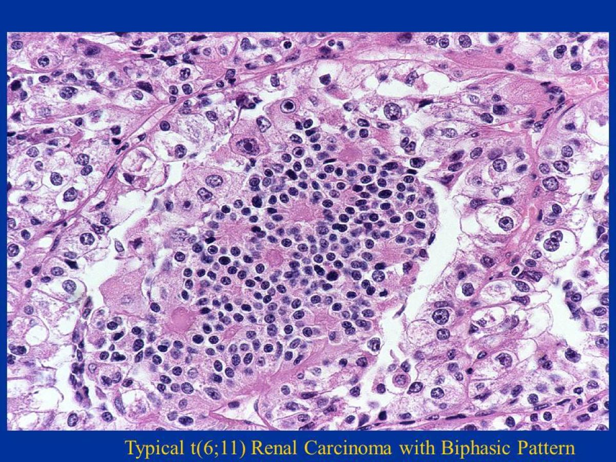
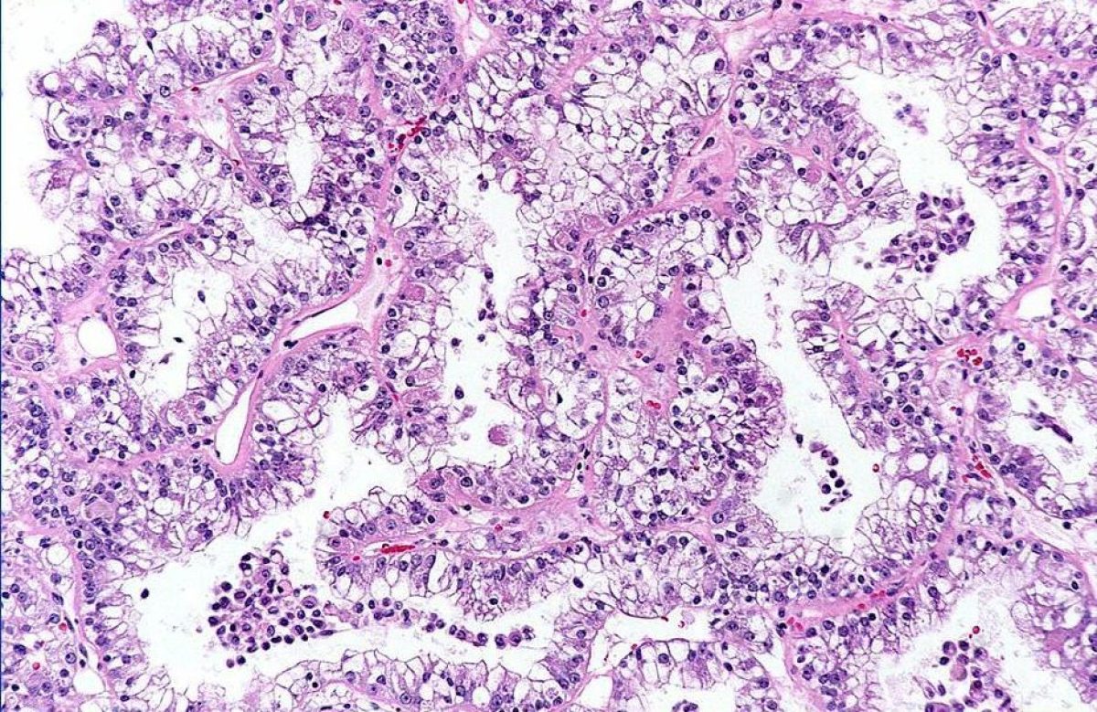
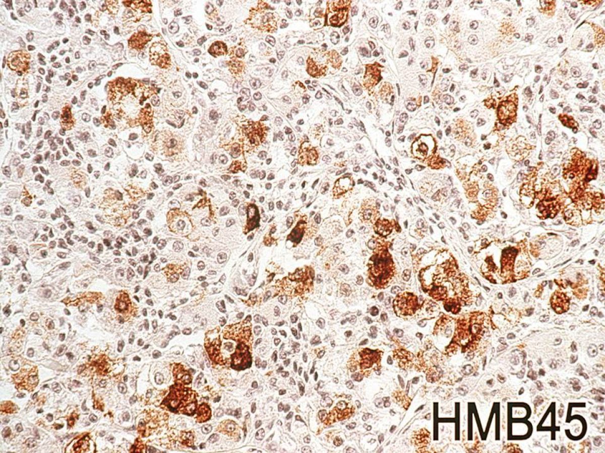
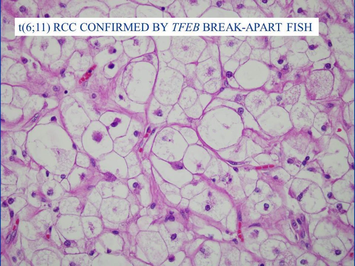
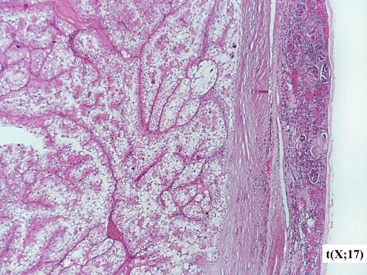
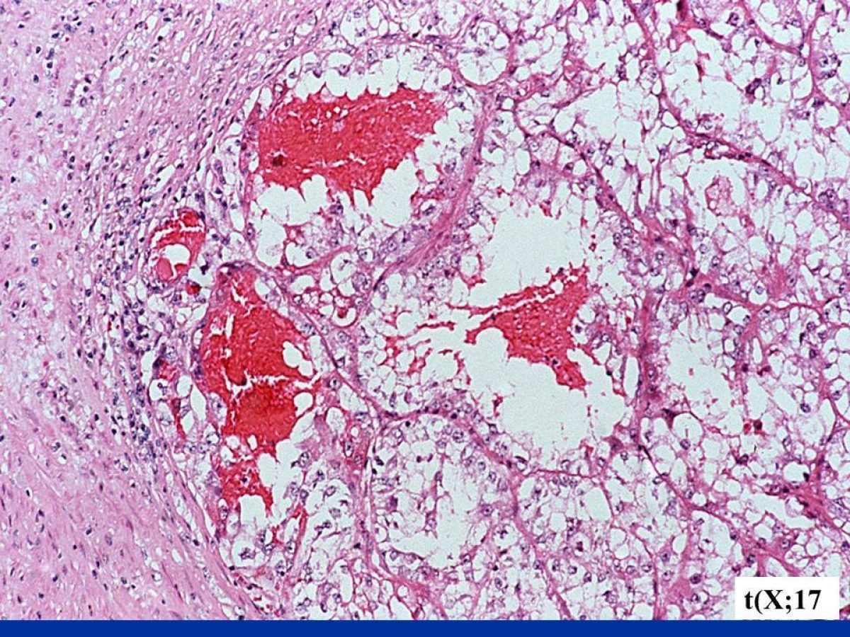
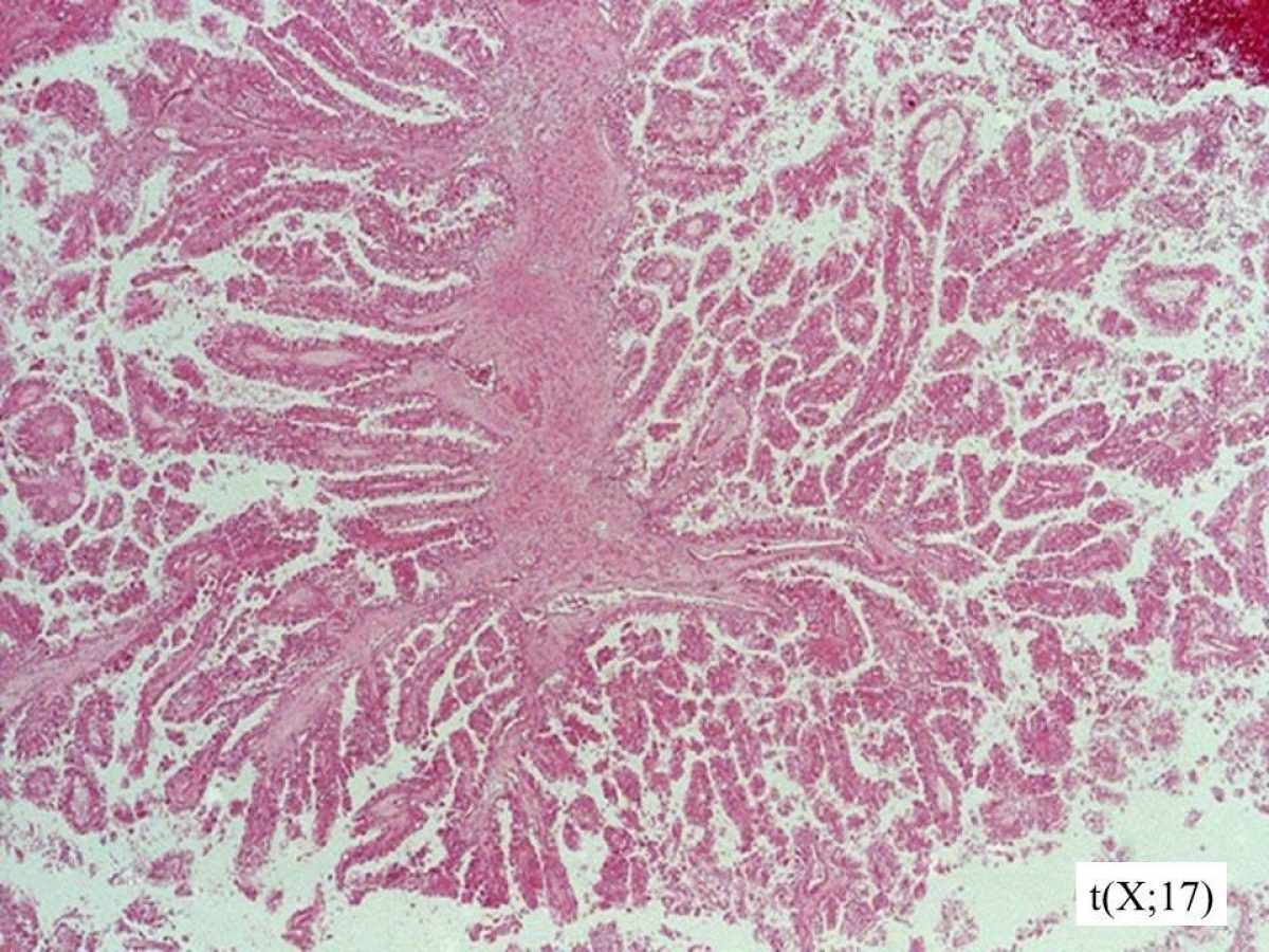
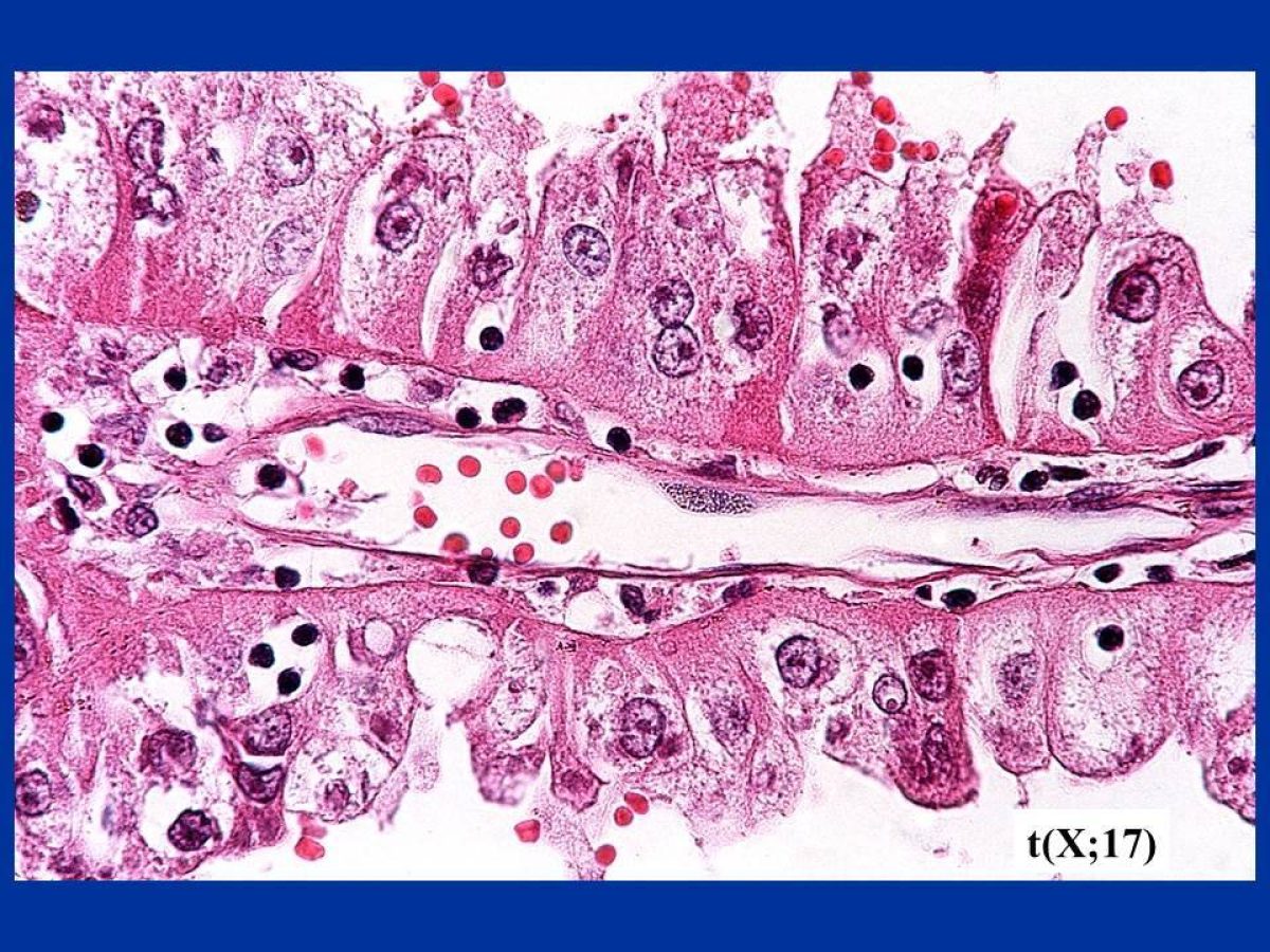
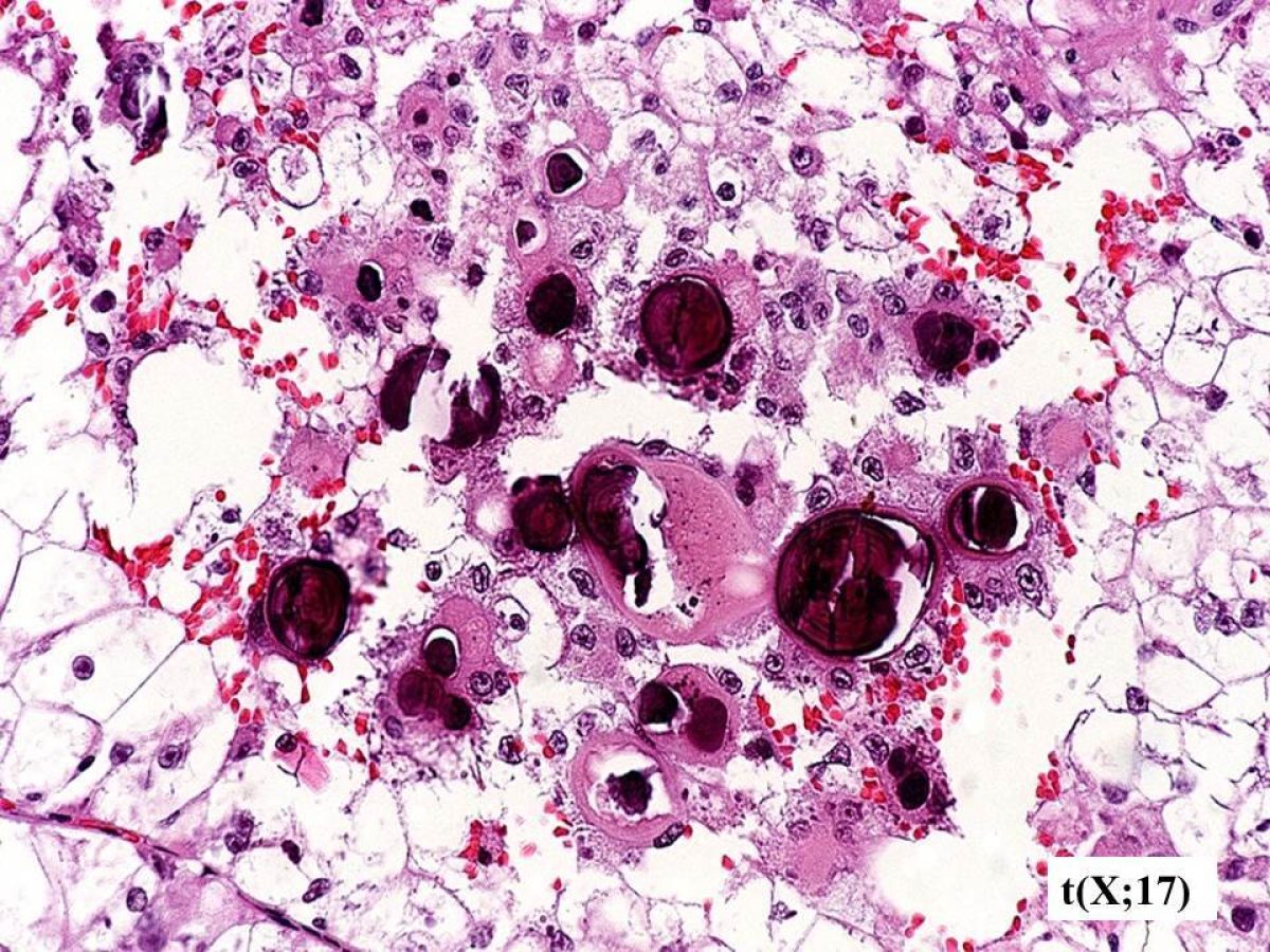
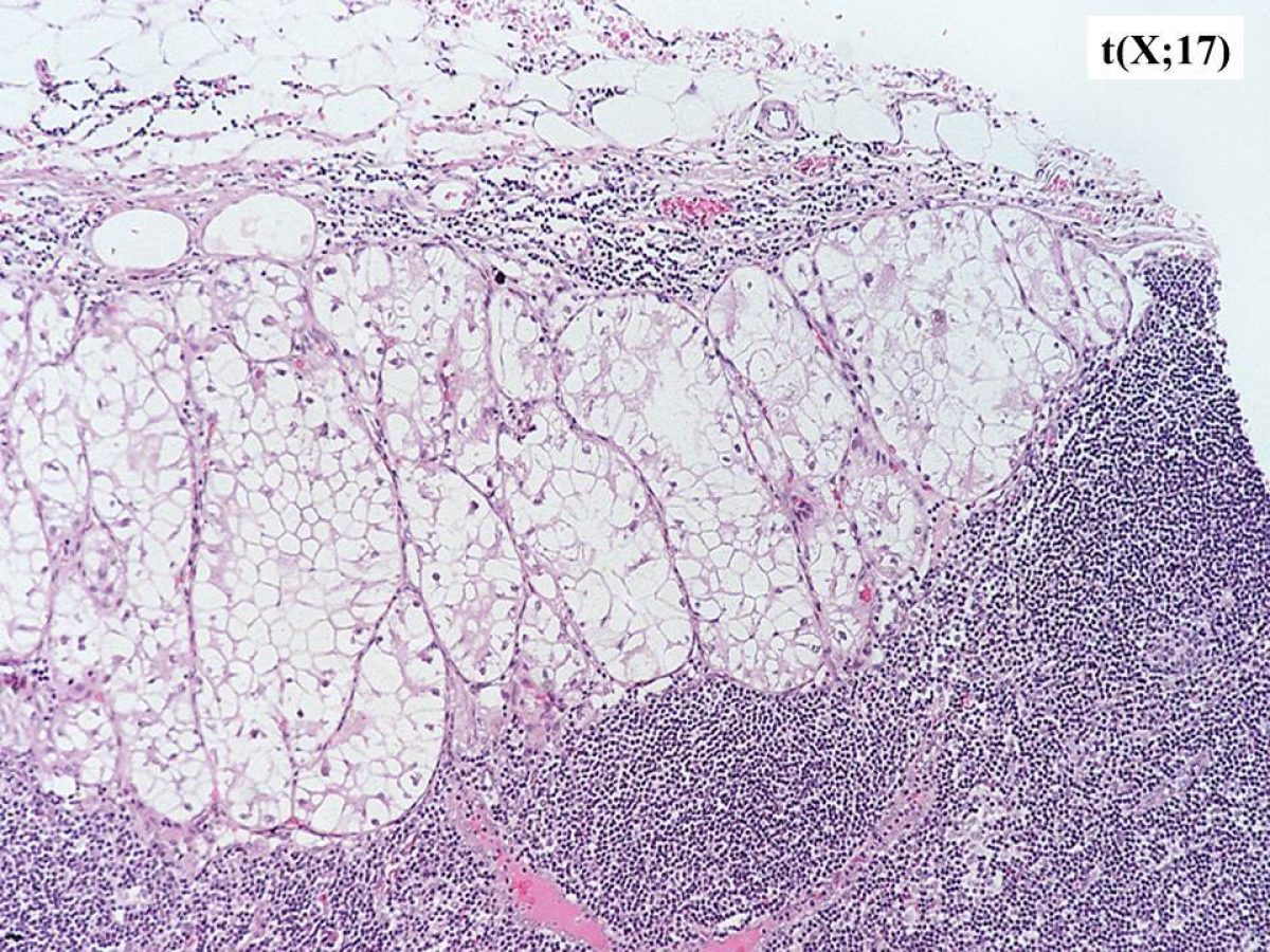
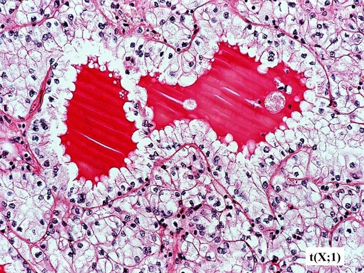
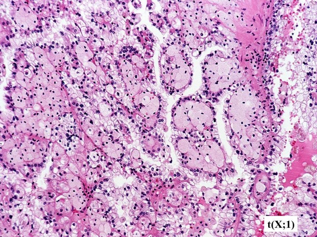
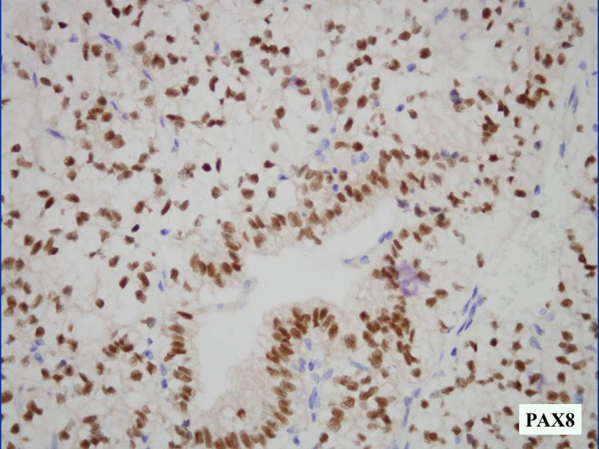
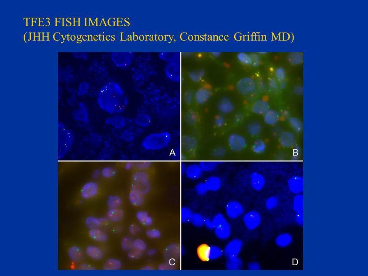
These pathology images are examples of what Translocation Renal Cell Carcinomas look like under the microscope.

TRCC Under the Microscope

  • pathology slide

    This is an Xp11 translocation renal cell carcinoma (RCC) with an ASPL-TFE3 gene fusion, resulting from a t(X;17) (p11;q25) chromosome translocation. This tumor has nested and papillary architecture, and contains clear cells.

  • pathology slide

    This ASPL-TFE3 RCC demonstrates prominent nested architecture and clear cells. Some Xp11 translocation RCC are mistaken for clear cell RCC.

  • pathology slide

    This ASPL-TFE3 RCC demonstrates prominent papillary architecture. Some Xp11 translocation RCC are mistaken for papillary RCC.

  • pathology slide

    This ASPL-TFE3 RCC demonstrates abundant, voluminous cytoplasm. The tumor cells are enormous compared to the adjacent red blood cells.

  • pathology slide

    The ASPL-TFE3 RCC often contain extensive psammomatous calcifications, which can make these tumors evident on plain X-rays of the abdomen.

  • pathology slide

    This ASPL-TFE3 RCC has metastasized to a lymph node. This subtype of Xp11 translocation RCC frequently spreads to lymph nodes; approximately 3 in 4 cases will have regional lymph node metastasis at diagnosis.

  • pathology slide

    The PRCC-TFE3 RCC result from a t(X;1) (p11;q21) chromosome translocation. These Xp11 translocation RCC also frequently show nested to acinar architecture, with blood or serum in the lumen. These tumors are frequently mistaken for clear cell RCC.

  • pathology slide

    This PRCC-TFE3 RCC demonstrates prominent papillary architecture with foam cells in the papillae, mimicking conventional papillary RCC.

  • pathology slide

    The Xp11 translocation RCC typical label for immunohistochemical markers of renal tubular origin, such as PAX8. In this case, the tumor shows strong nuclear labeling (brown), whereas the adjacent blood vessels are negative (blue).

  • pathology slide

    These images demonstrate tumors with evidence of TFE3 rearrangement by TFE3 FISH. Note the splitting of the red and green signals in the neoplastic cells.

  • pathology slide

    This is a t(6;11) (p21;q12) RCC. The tumor demonstrates a distinctive biphasic appearance, with larger clear cells and smaller cells clustered around pink nodules of basement membrane material.

  • pathology slide

    Some t(6;11) RCC can have clear cells and papillary areas that mimic the Xp11 translocation RCC.

  • pathology slide

    While the t(6;11) RCC are carcinomas, they characteristically demonstrate patchy immunolabeling for melanoma markers like HMB45.

  • pathology slide

    Some t(6;11) RCC look very similar to clear cell RCC, and thus the diagnosis can be missed unless there are clinical or pathologic clues to the diagnosis.

TRCC Under the Microscope

This is an Xp11 translocation renal cell carcinoma (RCC) with an ASPL-TFE3 gene fusion, resulting from a t(X;17) (p11;q25) chromosome translocation. This tumor has nested and papillary architecture, and contains clear cells.


This ASPL-TFE3 RCC demonstrates prominent nested architecture and clear cells. Some Xp11 translocation RCC are mistaken for clear cell RCC.


This ASPL-TFE3 RCC demonstrates prominent papillary architecture. Some Xp11 translocation RCC are mistaken for papillary RCC.


This ASPL-TFE3 RCC demonstrates abundant, voluminous cytoplasm. The tumor cells are enormous compared to the adjacent red blood cells.


The ASPL-TFE3 RCC often contain extensive psammomatous calcifications, which can make these tumors evident on plain X-rays of the abdomen.


This ASPL-TFE3 RCC has metastasized to a lymph node. This subtype of Xp11 translocation RCC frequently spreads to lymph nodes; approximately 3 in 4 cases will have regional lymph node metastasis at diagnosis.


The PRCC-TFE3 RCC result from a t(X;1) (p11;q21) chromosome translocation. These Xp11 translocation RCC also frequently show nested to acinar architecture, with blood or serum in the lumen. These tumors are frequently mistaken for clear cell RCC.


This PRCC-TFE3 RCC demonstrates prominent papillary architecture with foam cells in the papillae, mimicking conventional papillary RCC.


The Xp11 translocation RCC typical label for immunohistochemical markers of renal tubular origin, such as PAX8. In this case, the tumor shows strong nuclear labeling (brown), whereas the adjacent blood vessels are negative (blue).


These images demonstrate tumors with evidence of TFE3 rearrangement by TFE3 FISH. Note the splitting of the red and green signals in the neoplastic cells.


This is a t(6;11) (p21;q12) RCC. The tumor demonstrates a distinctive biphasic appearance, with larger clear cells and smaller cells clustered around pink nodules of basement membrane material.


Some t(6;11) RCC can have clear cells and papillary areas that mimic the Xp11 translocation RCC.


While the t(6;11) RCC are carcinomas, they characteristically demonstrate patchy immunolabeling for melanoma markers like HMB45.


Some t(6;11) RCC look very similar to clear cell RCC, and thus the diagnosis can be missed unless there are clinical or pathologic clues to the diagnosis.