Learn about Type 1 Diabetes development & progression under the microscope.

Type 1 Diabetes Pathology
Development of Type 1 Diabetes

The Pathology of Type 1 Diabetes

Insulin is a hormone the body produces to utilize the sugar in our bloodstream (absorbed from the food in our gut) as an energy source. In Type 1 Diabetes, insulin deficiency develops as the immune system attacks insulin producing cells as a result of inappropriately identifying them as foreign.

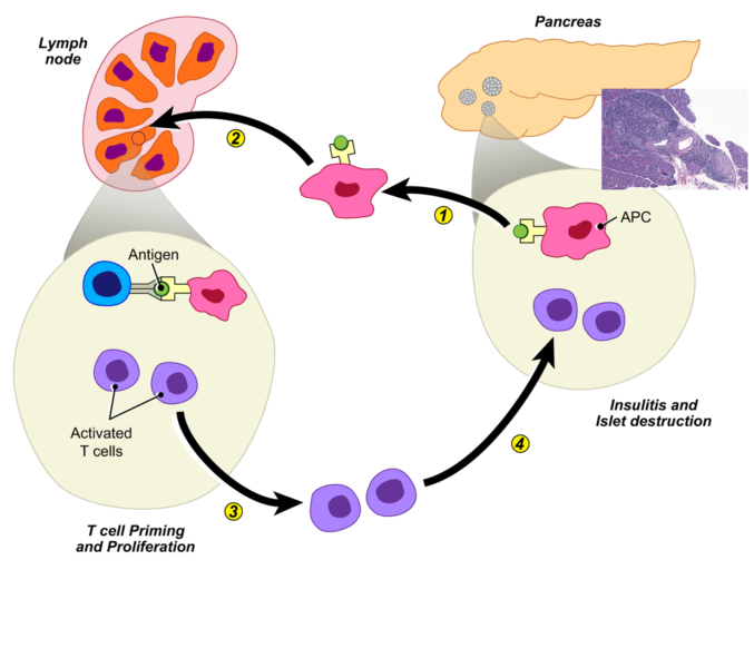
1) Antigen Presenting Cells (APCs) are exposed to insulin producing cells in the pancreas

2) These APCs travel to the lymph nodes and other parts of the body where to they involved in priming other parts of the immune system such as B Cells and T Cells against insulin-producing cells.

3) These B Cells and T Cells once activated, propagate this signal and proliferate.

4) These cells then search out and destroy insulin producing cells.

Without insulin, the sugar accumulates in the bloodstream and people can become very sick and present with very serious symptoms that include dehydration, weight loss, altered mental status, and even coma.

Meet Your Diabetes Experts

Meet Our Team
Insulitis
Insulitis: Infiltration of insulin-producing Beta Cells by pathogenic lymphocytes

Diagnosis of Type 1 Diabetes

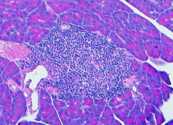
In order to diagnose someone with diabetes so they can receive appropriate treatment, we examine samples under the microscope to confirm the correct diagnosis (i.e., presence of certain antibodies) and differentiate Type 1 Diabetes from other conditions that may cause similar symptoms.

How Type 1 Diabetes Develops

  • Pancreas Pathology Slide

    A slide of the pancreas that houses pancreatic islets (cluster of cells) or Islets of Langerhans (1). These islets contain beta cells, which produce insulin.

  • Pancreas Pathology Slide

    A slide of a close-up on a healthy, insulin-producing, islet (1).

  • Pancreatic Pathology Slide

    A slide of pancreatic islets with a mix of healthy islets (1) and infiltrated cells (2), which occur as the lymphocytes (dark blue dots) of the immune system enter and destroy the cell.

  • Pancreatic Pathology Slide

    A slide of a close-up view of an infiltrated islet cell (2) being attacked by the immune system next to a blood vessel (1).

How Type 1 Diabetes Develops

A slide of the pancreas that houses pancreatic islets (cluster of cells) or Islets of Langerhans (1). These islets contain beta cells, which produce insulin.


A slide of a close-up on a healthy, insulin-producing, islet (1).


A slide of pancreatic islets with a mix of healthy islets (1) and infiltrated cells (2), which occur as the lymphocytes (dark blue dots) of the immune system enter and destroy the cell.


A slide of a close-up view of an infiltrated islet cell (2) being attacked by the immune system next to a blood vessel (1).