Our Division
As one of the premier cytopathology laboratories in the country, Hopkins Cytopathology plays a prominent role in the rapid, accurate diagnosis of the early stages of neoplastic lesions and infections and monitors disease recurrences.
Requests are welcomed from physicians seeking primary diagnoses, second opinions or more advanced services than they can provide, and from patients who self-refer for a rapid diagnosis or a second opinion. In addition to reviewing submitted slides and samples, our faculty pathologists can also extract cellular samples from patients who come to our clinic.
Experience
The Johns Hopkins Cytopathology Laboratory was one of the earliest cytopathology laboratories in the United States, established in 1956 by Dr. John K. Frost, a pioneer in the field. We have extensive experience serving practitioners throughout the United States, solving diagnostic dilemmas and providing second opinions. Gill's Hematoxylin, a widely used formula for staining the nuclei of cells for both cytopathology and histopathology specimens, was developed in the Hopkins Cytopathology Laboratory in the 1970s. Many other laboratory techniques developed here are described in literature accompanying various products and equipment, and in training films of the American Society of Cytopathology and The American Cancer Society. Today, the laboratory is directed by Dr. Syed Z. Ali
Expertise
Expert Consultation
Faculty who direct the laboratory are recognized experts in gynecologic, urologic, thyroid, pulmonary and pancreatic cytopathology, AIDS, and infectious disease pathology -- among other areas.
The majority of our cytotechnologists and cytopreparatory technicians who prepare specimens and review slides have at least 20 years experience in the field. This is an important advantage in an area that relies heavily on practiced judgment.
Special Capabilities and Services
Using a procedure developed at Hopkins and offered by few other labs, we can expand a single sample onto multiple different slides. Cell transfer technique enables us to get far more information from small samples, restore broken slides and create additional slides from a single slide for immunostaining techniques and for rare cases.
Fine needle aspiration of superficial lesions performed by an experienced cytopathologist offers the advantage of ensuring adequate specimen. Diagnoses can in some cases, be made immediately and appropriate patient management begun. Special studies such as cultures, flow cytometry and immuno stains, may be requested at the time of aspiration to avoid waiting until the specimen is sent to the laboratory for review. Sites lending themselves to this procedure are breast, lymph nodes, salivary gland, soft tissue lesions and thyroid gland.
We often work with Hopkins radiologists to extract deep tissue samples through endoscopy using a CT, MRI, or ultrasound to locate the site. Fine needle aspirations of deep organs, a minimally invasive outpatient procedure is less traumatic and less costly than exploratory surgery. Moreover, diagnosis can often be made on the spot so that appropriate patient management can begin immediately. Organs commonly aspirated are:
- thyroid gland
- liver
- lung
- all sites lymph nodes
- pancreas
Timely Results
When one of our pathologists obtains specimens from a patient, preliminary results can often be provided immediately. When we review submitted samples and slides, a written report and telephone consult can usually be provided within one or two days. Turnaround time for Pap test and other routine tests average three days.
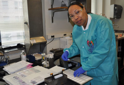
Technical staff
Cytotechnologists
Having over two centuries of experience in the field of cytotechnology, the nine Hopkins CTs bring a wealth of diagnostic expertise to their jobs every day. Working for less than a princely sum and having to pay handsomely for parking, there must be something intangible that this cadre of professionals possess. When asked why they work here and what they like about their jobs, they roll their collective eyes and usually give the age-old answer "collegiality, interesting patients and a varied, unusual caseload". Of course, there is one CT who occasionally states, "It's the only hospital within walking distance of my home!"
The JHH cytotechnologist's job description has changed over the fifty-four year history of The John K Frost Cytopathology Laboratory just as the field of cytotechnology has changed over that same time period. Gone are the days of conventional Pap smears and GI washes (although some of the staff still fondly remember them). Enter liquid-based gyn technology (absent any imaged cases at JHH), fine needle aspiration onsite evaluations, and molecular diagnostics. Most of the staff rotate turns in the endoscopy suite, neuroCT, and endocrine clinic, as well as an occasional trip to the OR. It is not particularly unusual for more CTs to be "out and about" with their carts than evaluating prepared specimens!
All this emphasis on diagnostic work is not to say that the Hopkins cytotechnologists are not involved in research activities. From the Early Lung Cancer Project in the early 70's through ALTS in the late 90's and now to FNA enhancement techniques, they collaborate with colleagues on papers and posters.
The varied workload, plus the collegiality, both intra- and extra-departmental makes working "at the Hop" stimulating and rewarding for this extraordinary group of professionals.