Pathology Department History
Gerald S. Spear
Written by Dr. Gerald S. Spear, M.D.
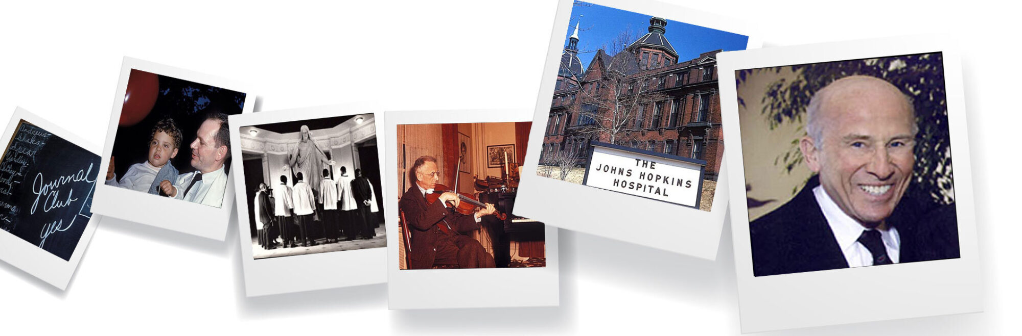
These photographs were taken by Dr. Gerald S. Spear JHMS 1952, House Staff Pathology 1953-1956, Chief Resident Pathology 1958-1959, Staff Pathology 1959-1977. The pictures were taken in 1955-1956 when he lived in the pathology resident's room on the 3rd floor in the south corridor under the Dome overlooking Broadway in what is now called the Billings Building. From 1956 to 1958 he was the founding Chief of the Histopathology Center at the 6407th USAF hospital at Tachikawa Japan. In 1977 Jerry left Hopkins to become Professor of Pathology at the University of California Irvine, retiring and becoming Emeritus in 2005. Upon his retirement the Department of Pathology at Irvine in collaboration with the Department of Pathology at Hopkins established a fellowship in his name for an Irvine student who excels in Pathology to participate in a one month elective in the Department of Pathology at Hopkins thereby to inspire respect for and, possibly, a career in pathology.
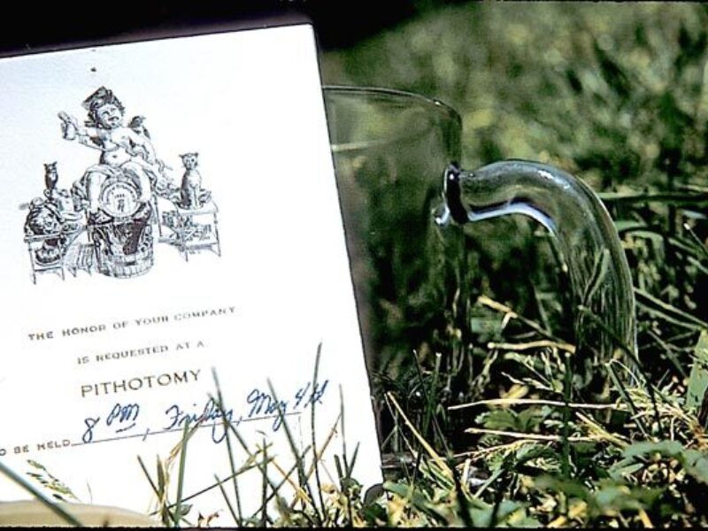
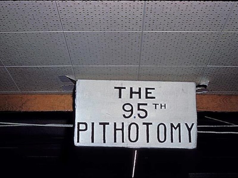
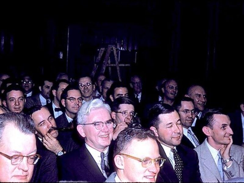
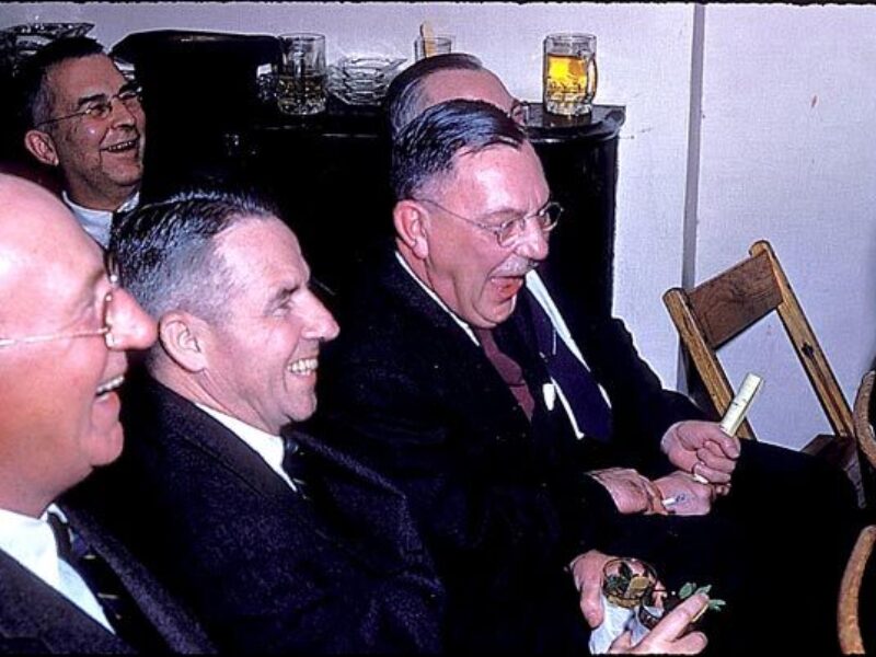
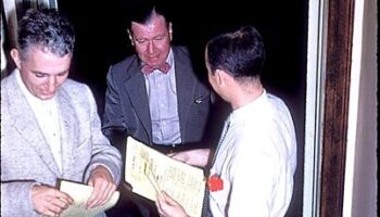
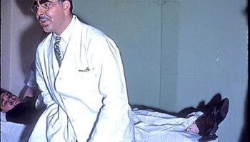
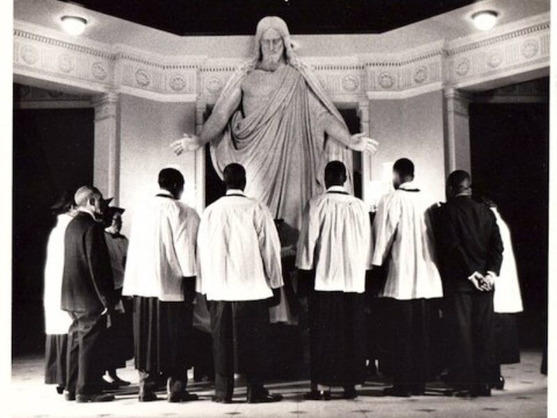
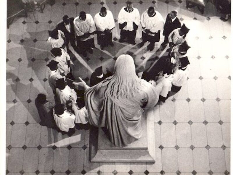
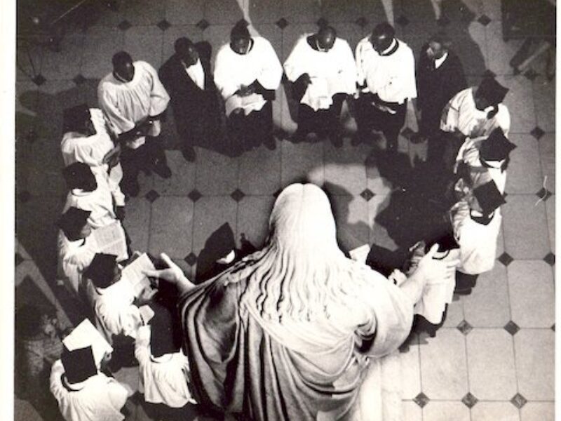
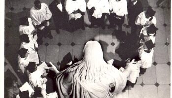
Below: The 95th Pithotomy Show May 1956
-
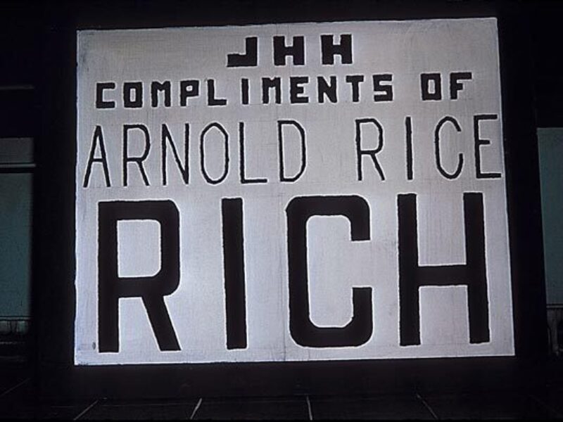
The 95th Pithotomy Show Photos (May 1956)
Pic 1 of 20
-
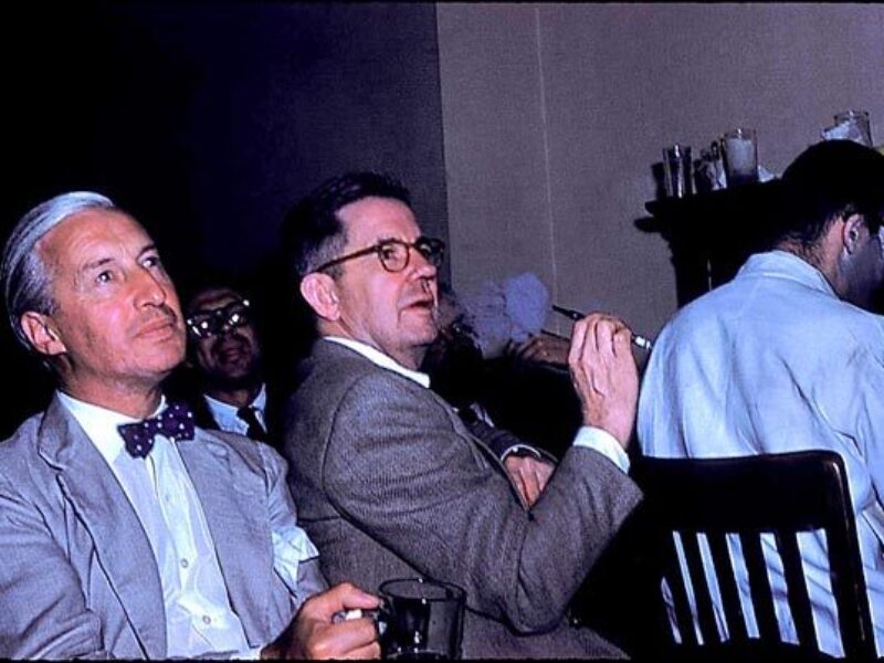
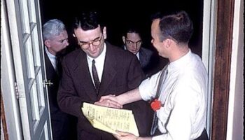
The 95th Pithotomy Show Photos (May 1956)
Pic 2 of 20
-
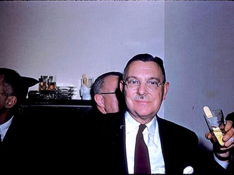
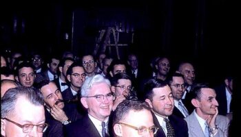
The 95th Pithotomy Show Photos (May 1956)
Pic 3 of 20
-
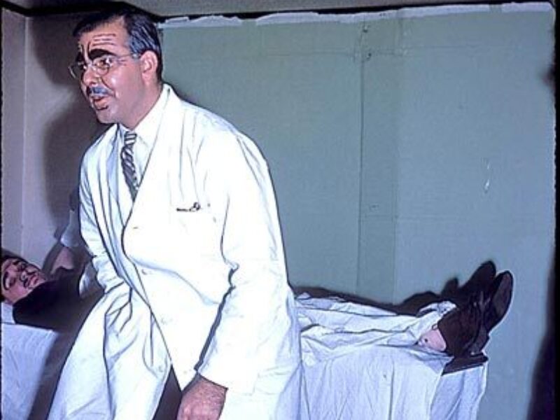
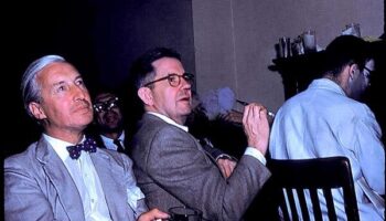
The 95th Pithotomy Show Photos (May 1956)
Pic 4 of 20
-
The 95th Pithotomy Show Photos (May 1956)
Pic 5 of 20
-
The 95th Pithotomy Show Photos (May 1956)
Pic 6 of 20
-
The 95th Pithotomy Show Photos (May 1956)
Pic 7 of 20
-
The 95th Pithotomy Show Photos (May 1956)
Pic 8 of 20
-
The 95th Pithotomy Show Photos (May 1956)
Pic 9 of 20
-
The 95th Pithotomy Show Photos (May 1956)
Pic 10 of 20
-
The 95th Pithotomy Show Photos (May 1956)
Pic 11 of 20
-
The 95th Pithotomy Show Photos (May 1956)
Pic 12 of 20
-
The 95th Pithotomy Show Photos (May 1956)
Pic 13 of 20
-
The 95th Pithotomy Show Photos (May 1956)
Pic 14 of 20
-
The 95th Pithotomy Show Photos (May 1956)
Pic 15 of 20
-
The 95th Pithotomy Show Photos (May 1956)
Pic 16 of 20
-
The 95th Pithotomy Show Photos (May 1956)
Pic 17 of 20
-
The 95th Pithotomy Show Photos (May 1956)
Pic 18 of 20
-
The 95th Pithotomy Show Photos (May 1956)
Pic 19 of 20
-
The 95th Pithotomy Show Photos (May 1956)
Pic 20 of 20
The 95th Pithotomy Show Photos (May 1956)
Pic 1 of 20
The 95th Pithotomy Show Photos (May 1956)

Pic 2 of 20
The 95th Pithotomy Show Photos (May 1956)

Pic 3 of 20
The 95th Pithotomy Show Photos (May 1956)
Pic 4 of 20
The 95th Pithotomy Show Photos (May 1956)
Pic 5 of 20
The 95th Pithotomy Show Photos (May 1956)
Pic 6 of 20
The 95th Pithotomy Show Photos (May 1956)
Pic 7 of 20
The 95th Pithotomy Show Photos (May 1956)
Pic 8 of 20
The 95th Pithotomy Show Photos (May 1956)

Pic 9 of 20
The 95th Pithotomy Show Photos (May 1956)
Pic 10 of 20
The 95th Pithotomy Show Photos (May 1956)
Pic 11 of 20
The 95th Pithotomy Show Photos (May 1956)

Pic 12 of 20
The 95th Pithotomy Show Photos (May 1956)

Pic 13 of 20
The 95th Pithotomy Show Photos (May 1956)
Pic 14 of 20
The 95th Pithotomy Show Photos (May 1956)

Pic 15 of 20
The 95th Pithotomy Show Photos (May 1956)

Pic 16 of 20
The 95th Pithotomy Show Photos (May 1956)
Pic 17 of 20
The 95th Pithotomy Show Photos (May 1956)
Pic 18 of 20
The 95th Pithotomy Show Photos (May 1956)

Pic 19 of 20
The 95th Pithotomy Show Photos (May 1956)
Pic 20 of 20
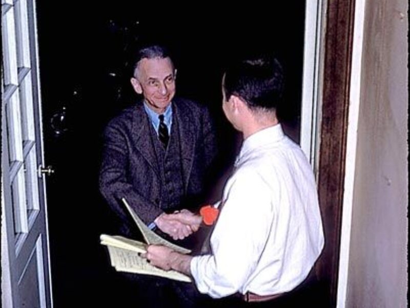
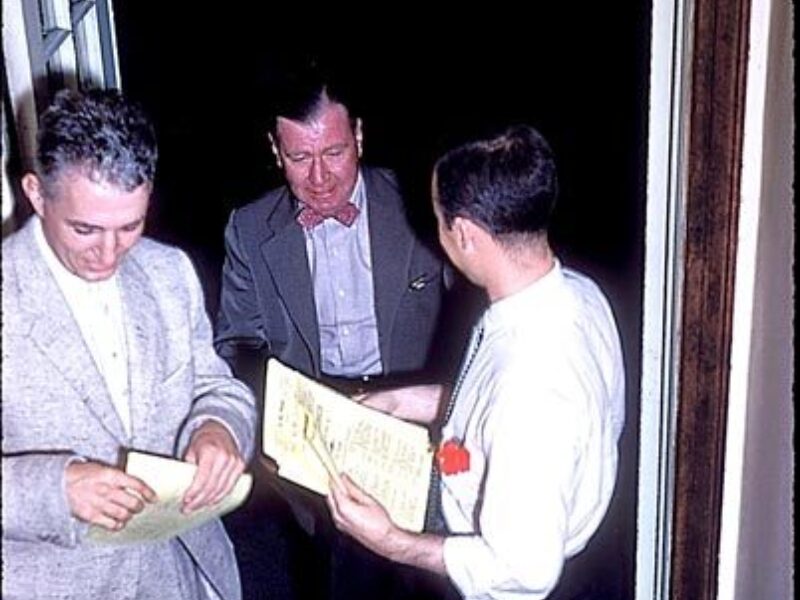
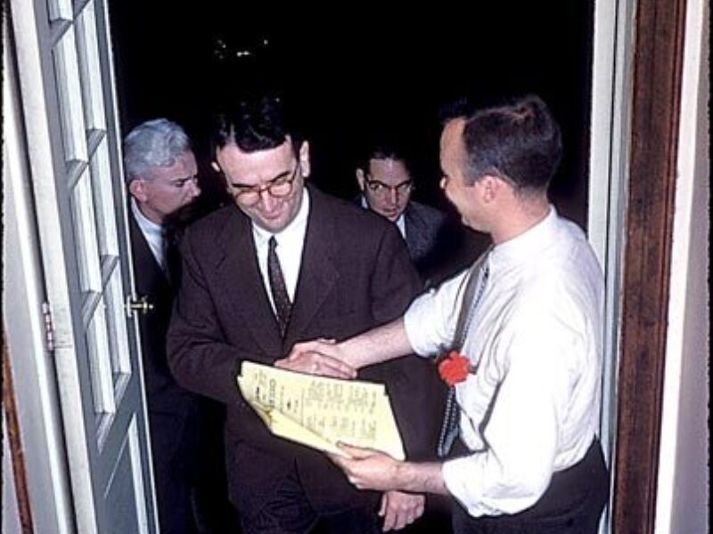
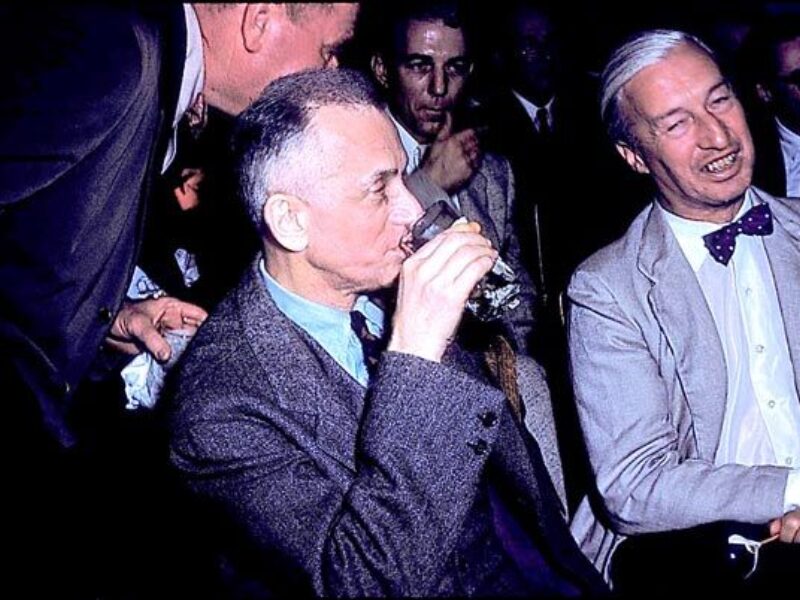
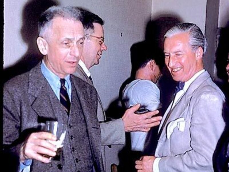
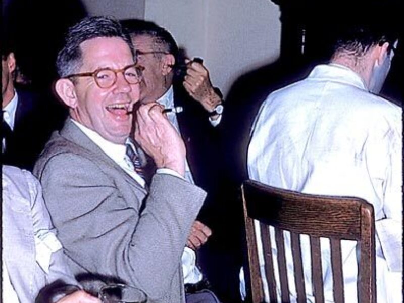
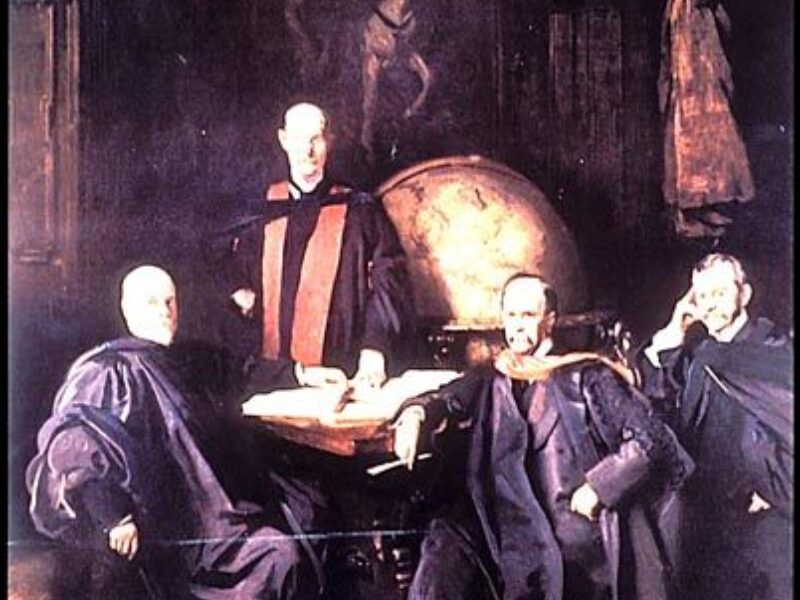
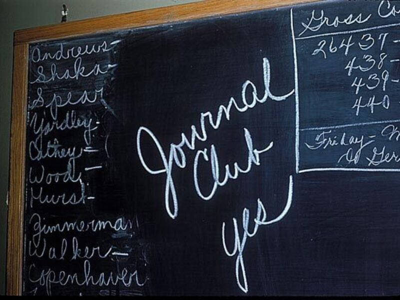
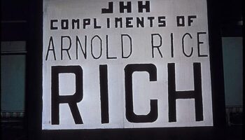
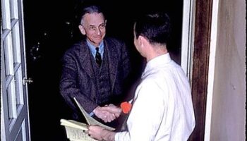
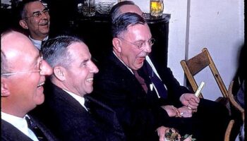
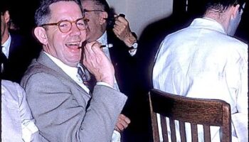
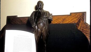
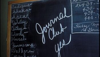
Journal Club at home of Dr. and Mrs. Rich likely in May 1956
Turtle Derby
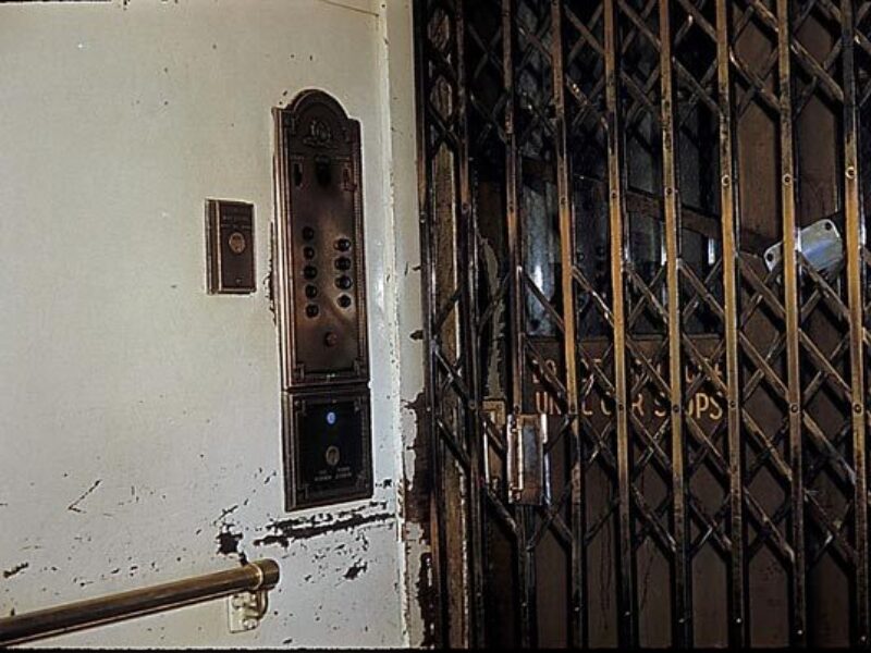
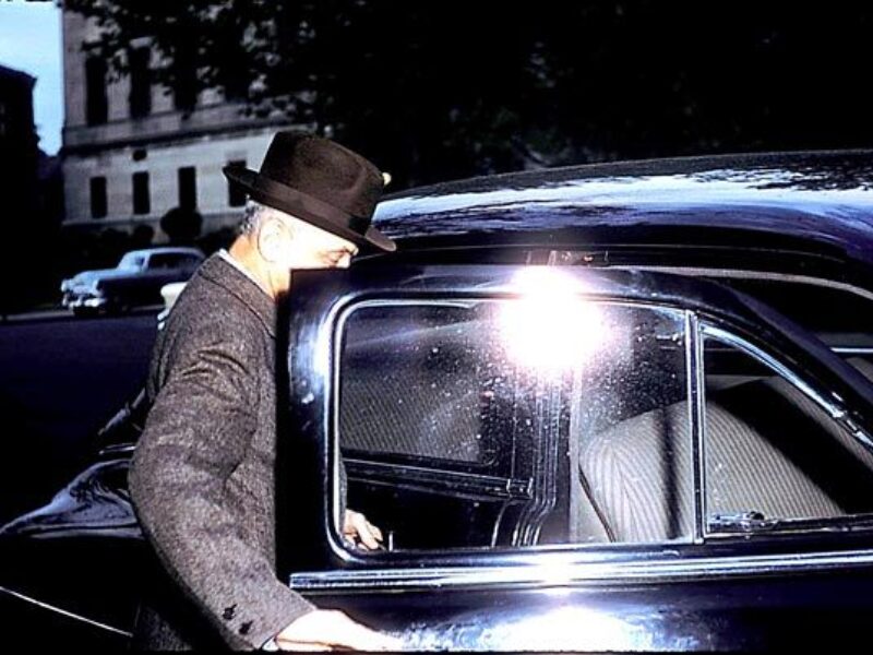
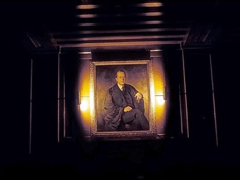
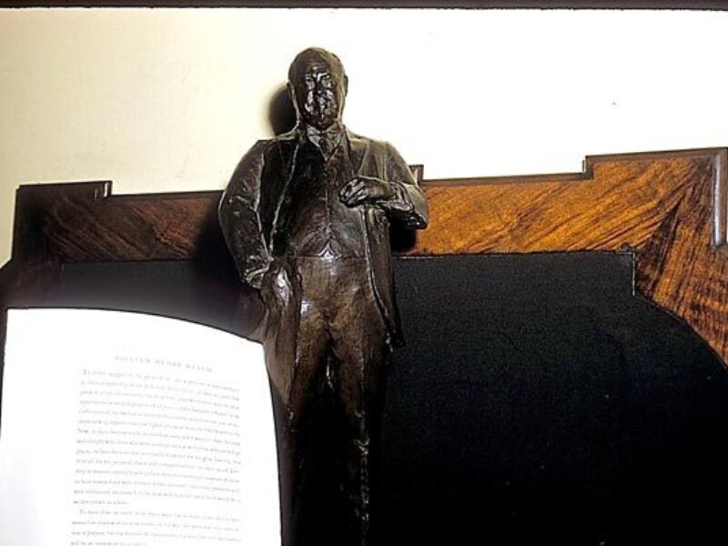
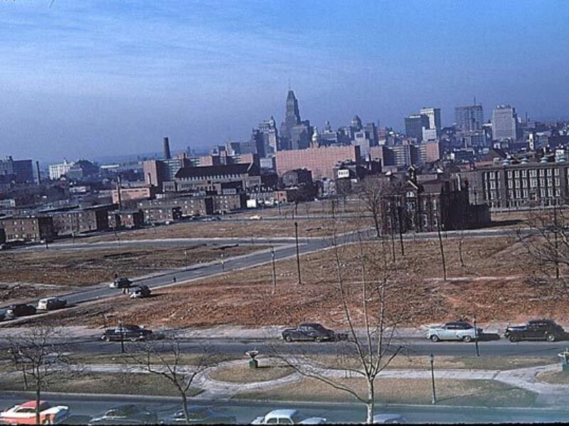
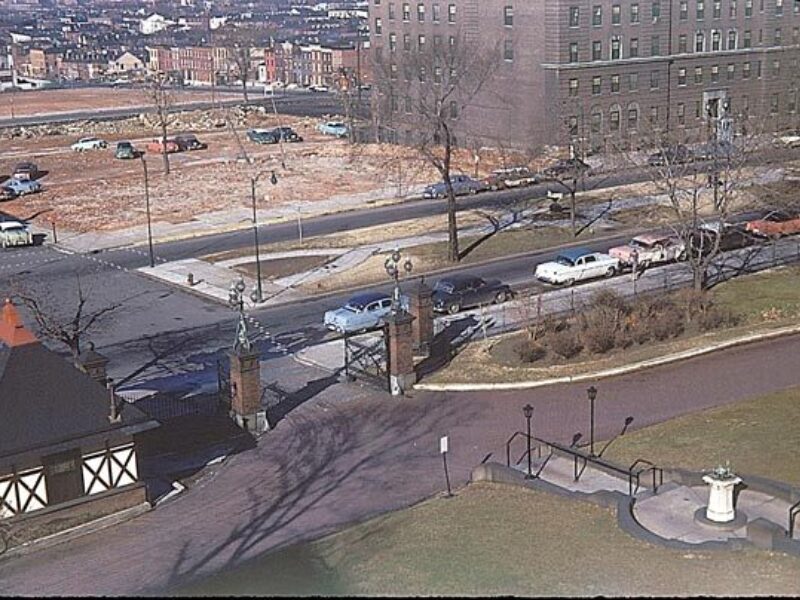
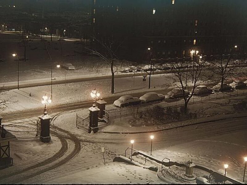
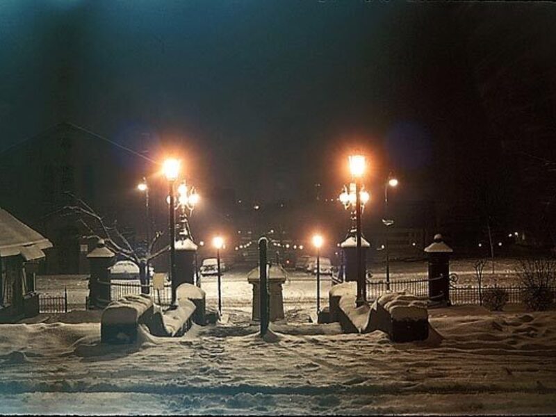
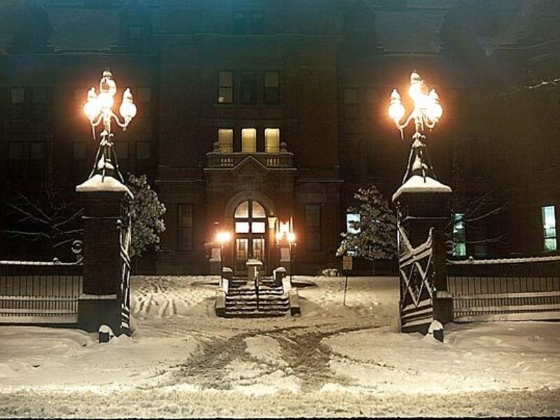
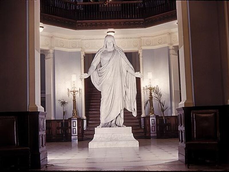
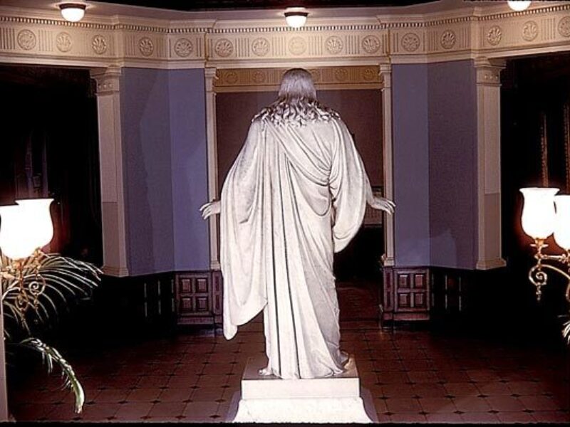
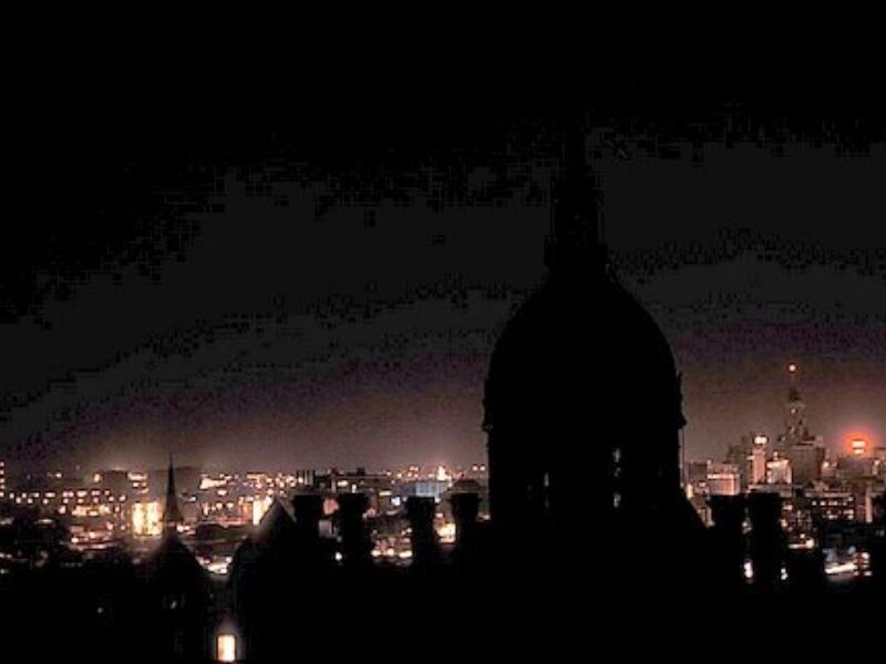
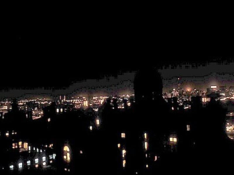
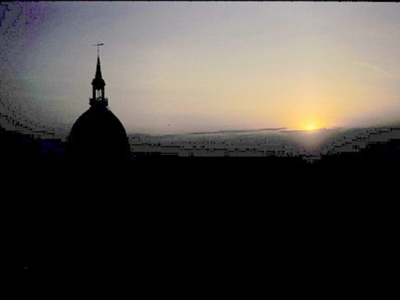
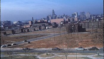
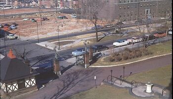
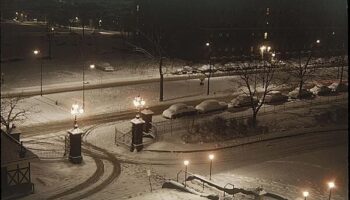
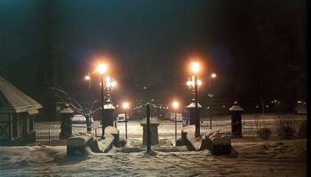
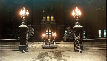
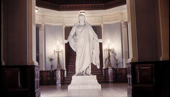
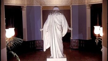
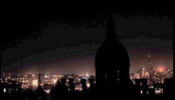
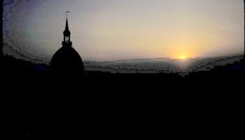
Below: Views of and from the Hospital during the period in my residency when I lived in the Pathology Quarters (i.e. room) that was reserved for and assigned to Pathology Residents on the third floor of the front Administration Building of the Hospital in the south wing, the room, being on the west side of the corridor, fronting on Broadway. The room may have been the room with the bay window. These pictures were taken in the winter of 1955-56. I was the only Pathology Resident living in the Hospital under this arrangement at the time. In fact, 1955-56 may have been the last year that residents lived in the administration building. Rooms transitioned to administrative staff offices, and, in fact, I believe this transition was occurring as I lived there.
-
Hospital Photos
Pic 1 of 13
-
Hospital Photos
Pic 2 of 13
-
Hospital Photos
Pic 3 of 13
-
Hospital Photos
Pic 4 of 13
-
Hospital Photos
Pic 5 of 13
-
Hospital Photos
Pic 6 of 13
-
Hospital Photos
Pic 7 of 13
-
Hospital Photos
Pic 8 of 13
-
Hospital Photos
Pic 9 of 13
-
Hospital Photos
Pic 10 of 13
-
Hospital Photos
Pic 11 of 13
-
Hospital Photos
Pic 12 of 13
-
Hospital Photos
Pic 13 of 13
Hospital Photos
Pic 1 of 13
Hospital Photos
Pic 2 of 13
Hospital Photos
Pic 3 of 13
Hospital Photos
Pic 4 of 13
Hospital Photos
Pic 5 of 13
Hospital Photos
Pic 6 of 13
Hospital Photos
Pic 7 of 13
Hospital Photos
Pic 8 of 13
Hospital Photos

Pic 9 of 13
Hospital Photos
Pic 10 of 13
Hospital Photos

Pic 11 of 13
Hospital Photos

Pic 12 of 13
Hospital Photos
Pic 13 of 13
Brief History of Jerry Spear's many Contributions to Renal Biopsy at Johns Hopkins
Gerald ("Jerry") Spear contributed significantly to the introduction of renal pathology at Johns Hopkins. Below is a brief timeline that highlight Jerry's impact.
| Year | Jerry's contributions |
|---|---|
| 1950 | Coons and Kaplan from Harvard report immunofluorescent microscopic detection of tissue bound immune deposits. |
| 1955 | Mellors First application of the technique to renal tissue. |
| 1960 | Jerry Spear's first publication on renal pathology, a light microscopic autopsy study of the glomeruli in cyanotic congenital heart disease and primary pulmonary hypertension recognizing distinctive glomerular lesions. (1) |
| July 1960 - July 1962 | Robert Heptinstall is a visiting Professor of Pathology at Washington University St. Louis |
| Early 1960's (or earlier) - 1976 | Jerry Spear was responsible for examination and reporting of percutaneous renal biopsies at Johns Hopkins being one of the early institutions utilizing light, electron as well as immunofluorescent microscopy. |
| 1962 | Robert H. Heptinstall came to Hopkins Department of Pathology concentrating for some time on the preparation of the first edition of his book, 'Pathology of the Kidney', which is published in 1966. |
| 1964 | Jerry Spear's first immunofluorescent publication. This was the first report in human kidney of what became the classic diffuse mesangial pattern of immunofluorescence. (2) |
| 1965 | Jerry Spear's first renal biopsy manuscript being an overview of expectatioms from percutaneous renal biopsy based on personal experience during the prior years.(3) |
| October 1966 - June 1969 | Heptinstall appointed Acting Director and Pathologist in Charge Department of Pathology. |
| 1969 - 1988 | Heptinstall was Baxley Professor, Director, Pathologist in Chief Johns Hopkins University School of Medicine and Hospital. |
| 1970 | Jerry Spear's first immunofluorescent manuscript on percutaneous renal biopsies being a study of Alport Syndrome demonstrating discordant immunoglobulin-complement reactions with first speculation therefore that immune complexes may not play a role in hereditary nephritis. (4) |
| 1971 | Jerry Spear authored two papers describing renal biopsy findings in cystinosis, the first (5) a unique ultrastructural and electron probe study, the second (6) highlighting distinctive polykaryocytosis of the visceral glomerular epithelium and emphasizing the important diagnostic significance of the latter. |
| 1971 | In this paper (7) Spear and colleagues describe the clinical and pathological biopsy findings in an infant and suggest that pseudohermaphroditism, chronic glomerulonephritis and Wilms' tumor appear to constitute a clinical syndrome which commonly is designated as Drash syndrome. |
| 1972 | In this paper (8) Spear described a distinctive ultrastructural lesion in glomerular basement membrane in Alport Syndrome permitting diagnosis and playing a role in understanding its molecular pathogenesis. Another group reported the same lesion in the same year, these being the first descriptions in the medical literature. |
| 1973 | Jerry Spear proposed a theory (9) for the pathogenesis of Alport Syndrome based on the above (8) and upon genetic and biochemical principles namely the basis of the syndrome may be heterogeneous abnormalities in a gene or genes encoding basement membrane collagen in the glomerulus, lens capsule and inner ear, possibly in some instances effecting replacement or deletion of half-cystine. This theory has been commented upon by Kashtan (10). |
| 1973 | In this paper (11) studying idiopathic hematuria of childhood Spear pointed out that the ultrastructural glomerular lesions characteristic of Alport syndrome were not present a finding which may permit differentiation of these two types of hematuria in children which is often a difficult clinical and pathological problem. |
| 1973 | In this paper (12) Spear pointed out that contrary to usual belief little if any visible evidence exists to support the notion that the swan-neck lesion and Fanconi syndrome in cystinosis are a result of the accumulation of cystine in the epithelium of the proximal tubule. |
References
- Glomerular alterations in cyanotic congenital heart disease. Spear GS. Bull. Johns Hopkins Hosp. 1960 Jun;106:347-67.
- The glomerulus in cyanotic congenital heart disease:an immunofluorescent study. Spear GS, Kihara I. Bull. Johns Hopkins Hosp. 1964 Dec;115:481-93.
- Conjoint clinic on renal biopsy: perspective and clinicopathological considerations. Spear GS. J Chronic Dis. 1965 Feb;18:133-145.
- Hereditary nephritis with nerve deafness. Immunofluorescent studies on the kidney, with a consideration of discordant immunofluorescent reactions. Spear GS, Whitworth JM, Konigsmark BW. Am J Med. 1970 Jul;49(1):52-63.
- Cystinosis. An ultastructural and electron-probe study of the kidney with unusual findings, Spear GS, Slusser RJ, Tousimis AJ, Taylor CG, Schulman JD. Arch Pathol. 1971 Mar;91 (3):206-21.
- Polykaryocytosis of the visceral glomerular epithelium in cystinosis with a description of an unusual clinical variant. Spear GS, Slusser RJ. Schulman JD, Alexander F. Johns Hopkins Med J. 1971 Aug;129(2):83-99.
- Pseudohermaphroditism, glomerulonephritis with the nephrotic syndrome, and Wilms' tumor in infancy. Spear GS, Hyde TP, Gruppo RA, Slusser R.j. J Pediatr. 1971 Oct;79(4):677-81.
- Alport's syndrome. Emphasizing electron microscopic studies of the glomerulus. Spear GS, Slusser RJ. Am J Pathol. 1972 Nov;69(2):213-24.
- Editorial: Alport's Syndrome: a consideration of pathogenesis. Spear GS. Clin Nephrol. 1973 Nov-Dec; 1(6):336-7.
- Alport Syndrome. Kashtan CE, Michael AF. Kid Int 1996 (50): 1445-1463.
- Idiopathic hematuria of childhood. Pathologic findings in the kidney in six patients. Spear GS, Roskes SD, Slusser RJ, Alsruhe JP. Hum Pathol. 1973 Sep;4(3): 349-80.
- The proximal tubule and the podocyte in cystinosis. Spear G. Nephron 1973 (10):57-60.